
Aging reorganizes activity across the whole brain.
Whole-brain c-Fos mapping in young and aged mice reveals a systematic redistribution of resting brain activity — and powers Theracast’s Brain Aging Platform: quantifying aging, and the effect of interventions on it, region by region.
A forebrain-to-hindbrain rebalancing.
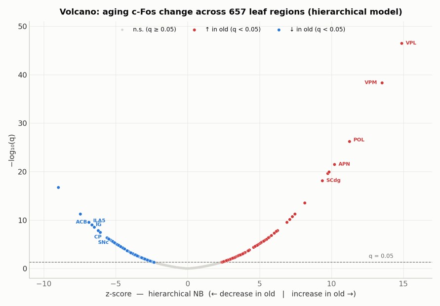
Aging does not change c-Fos+ cell distribution uniformly — it redistributes it along a clear forebrain-to-hindbrain axis. Two complementary statistical models agree: of 657 anatomical regions, 252 differ significantly between young and old animals.
Dominate the forebrain: medial prefrontal and cingulate cortex, striatum and pallidum, accumbens, septum, olfactory cortex, and the dopaminergic and noradrenergic nuclei.
Dominate the sensory and subcortical brain: relay thalamus, superior colliculus, reticular midbrain, and cerebellar cortex.
Aging rewires brain-network coupling.
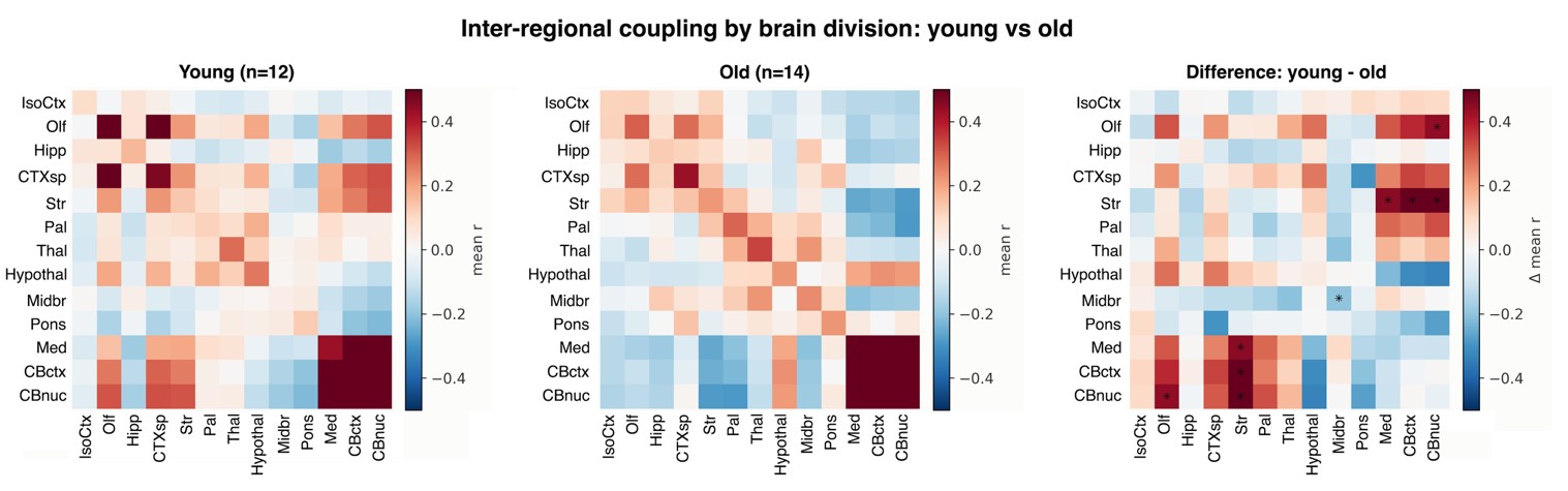
How brain regions co-vary across animals is itself reorganized (p < 0.001). Overall coupling strength is unchanged — but coupling is preserved within brain systems while collapsing between them: old brains are more de-integrated across systems. The forebrain specifically decouples from the cerebellum and medulla, reversing from positive to negative coupling.
A top-down to bottom-up rebalancing.
Anatomically, forebrain executive and limbic control systems — medial prefrontal/cingulate cortex, the cortico-striatal-pallidal loops, and the dopaminergic and noradrenergic nuclei that drive them — lose c-Fos-based activity, while sensory, orienting, and cerebellar systems gain it. At the network level, coupling stays preserved locally but weakens between systems.
The mouse findings parallel human brain aging.
Frontal aging & default-mode decline
The prefrontal/cingulate decrease agrees with the frontal-aging hypothesis and default-mode-network decline — the rodent counterparts of those human networks.
Striatal dopamine loss
The substantia nigra/VTA and striatum decrease matches the well-documented loss of striatal dopamine with age.
Hippocampal shift
Dentate/entorhinal-input decrease with CA1/subicular-output increase mirrors the human shift toward CA hyperactivity and dentate hypofunction.
Neural dedifferentiation
The amplification of sensory systems parallels dedifferentiation seen in human aging.
System segregation
The coupling reorganization connects to human work on age-related changes in the segregation of large-scale brain systems.
Why it matters
A mouse assay that recapitulates human aging signatures can screen interventions — and read out their brain-wide effects in one experiment.
Quantify brain aging — and what your intervention does to it.
The same platform that profiles psychiatric drugs measures aging-related change across every brain region in a single assay, in absolute counts and brain-wide shares.
Contact Pavel Osten